- GET CORRECTLY DIAGNOSED NOW - WORLD CLASS EMG TESTING
- 312-877-0035
EMG Best Test for Cervical Radiculopathy (Pinched Nerves)
Cervical radiculopathy, commonly called a “pinched nerve” occurs when a nerve in the neck is compressed or irritated where it branches away from the spinal cord.
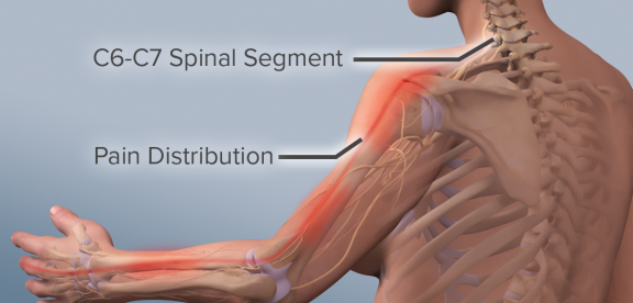
Cervical radiculopathy may cause pain that radiates into the shoulder, as well as muscle weakness and numbness that travels down the arm and into the hand.
What is Cervical Radiculopathy
Cervical radiculopathy is often caused by “wear and tear” changes that occur in the spine as we age, such as arthritis. When related to trauma or Injury, it is most often caused by a sudden movement that results in a herniated disk, torn ligaments, the development of scar tissue blocking a canal or nerve track or exit zone.
In most cases, cervical radiculopathy responds well to conservative treatment that includes medication and physical therapy.
Anatomy
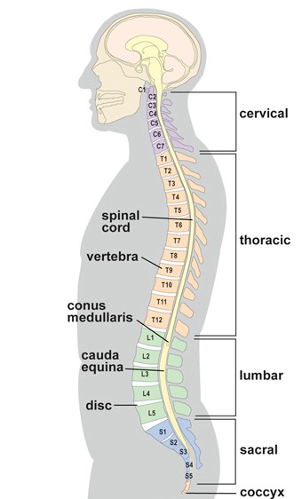
Your spine is made up of 24 bones, called vertebrae, that are stacked on top of one another. These bones connect to create a canal that protects the spinal cord. The seven small vertebrae that begin at the base of the skull and form the neck comprise the cervical spine.
Cervical radiculopathy occurs in the cervical spine — the seven small vertebrae that form the neck.
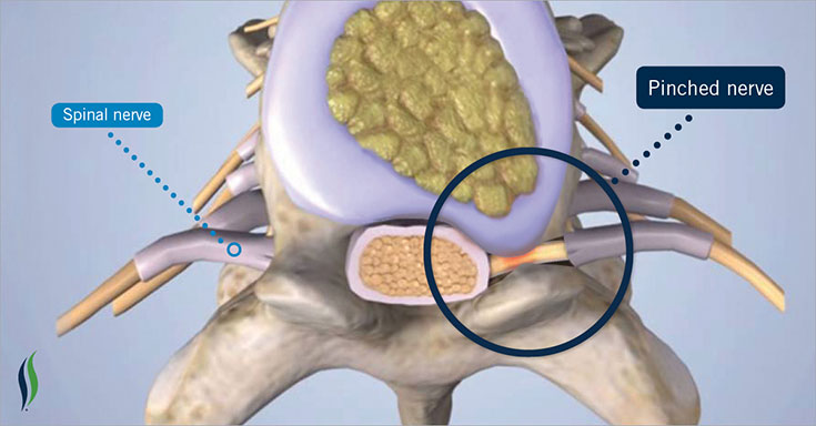
Other parts of your spine include your spinal cord and nerves. These “electrical cables” travel through the spinal canal carrying messages between your brain and muscles. Nerve roots branch out from the spinal cord through openings in the vertebrae (foramen).

An intervertebral disk Herniation (cross-section view)
Intervertrebral disks. In between your vertebrae are flexible intervertebral disks. They act as shock absorbers when you walk or run. Intervertebral disks are flat and round and about a half inch thick.
They are made up of two components:
Annulus fibrosus. This is the tough, flexible outer ring of the disk.
Nucleus pulposus. This is the soft, jelly-like center of the disk.
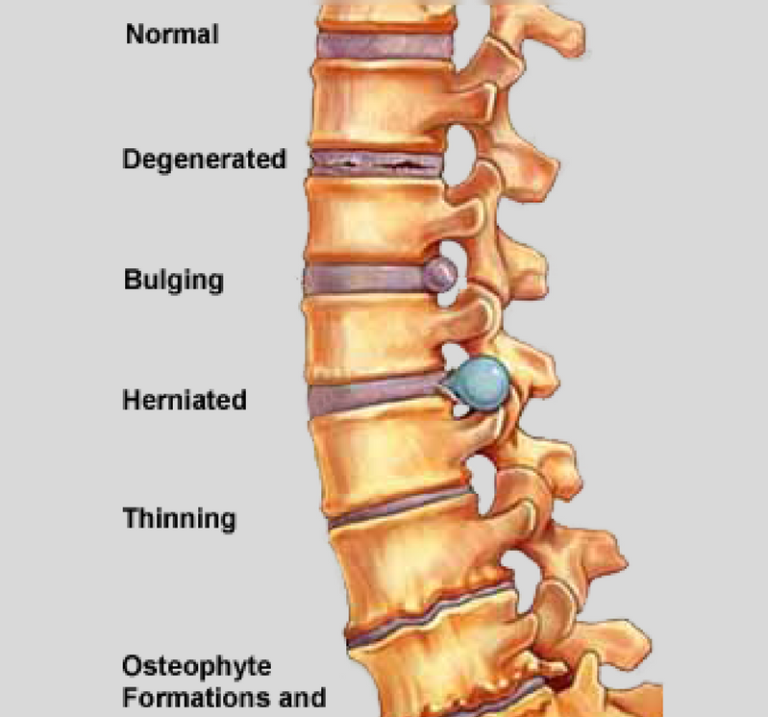
Cervical radiculopathy most often arises from degenerative changes that occur in the spine as we age or from an injury that causes a herniated, or bulging, intervertebral disk.
The Cause of Pinched Nerves
Degenerative changes. As the disks in the spine age, they lose height and begin to bulge. They also lose water content, begin to dry out, and become stiffer. This problem causes settling, or collapse, of the disk spaces and loss of disk space height.
As the disks lose height, the vertebrae move closer together. The body responds to the collapsed disk by forming more bone —called bone spurs—around the disk to strengthen it. These bone spurs contribute to the stiffening of the spine. They may also narrow the foramen—the small openings on each side of the spinal column where the nerve roots exit—and pinch the nerve root.
Degenerative changes in the disks are often called arthritis or spondylosis. These changes are normal and they occur in everyone. In fact, nearly half of all people middle-aged and older have worn disks and pinched nerves that do not cause painful symptoms. It is not known why some patients develop symptoms and others do not.
Herniated disk. A disk herniates when its jelly-like center (nucleus) pushes against its outer ring (annulus). If the disk is very worn or injured, the nucleus may squeeze all the way through. When the herniated disk bulges out toward the spinal canal, it puts pressure on the sensitive nerve root, causing pain and weakness in the area the nerve supplies.
A herniated disk often occurs with lifting, pulling, bending, or twisting movements.
In a herniated disk, the soft, jelly-like center of the disk can push all the way through the outer ring. (Side and cross-section views shown)
Symptoms
In most cases, the pain of cervical radiculopathy starts at the neck and travels down the arm in the area served by the damaged nerve. This pain is usually described as burning or sharp. Certain neck movements—like extending or straining the neck or turning the head—may increase the pain.
Other symptoms include:
- Tingling or the feeling of “pins and needles” in the fingers or hand
- Weakness in the muscles of the arm, shoulder, or hand
- Loss of sensation
Some patients report that pain decreases when they place their hands on top of their head. This movement may temporarily relieve pressure on the nerve root.
YOUR DOCTOR EXAMINATION
Physical Examination
After discussing your medical history and general health, your doctor will ask you about your symptoms. He or she will then examine your neck, shoulder, arms and hands—looking for muscle weakness, loss of sensation, or any change in your reflexes. Your doctor may also ask you to perform certain neck and arm movements to try to recreate and/or relieve your symptoms.
Tests
Electromyography (EMG). Electromyography measures the electrical impulses of the muscles at rest and during contractions. Nerve conduction velocity studies are often done along with EMG to determine if a nerve is functioning normally. Together, these tests are the MOST IMPORTANT DIAGNOSTIC TEST you can get to determine nerve dysfunction or damage. They will help your doctor determine whether your symptoms are caused by pressure on spinal nerve roots and nerve damage or by another condition that causes damage to nerves, such as diabetes. When Pain Management Physicians, Orthopedic or Spine Surgeons order these tests, they order them to identify exactly where you have nerve compression, irritation, swelling or reduced or non-conduction of the nerve. This information is extremely important and allows them to choose the correct Therapy or Procedure to restore your health and minimize your pain.
X-rays. These provide images of dense structures, such as bone. An x-ray will show the alignment of bones along your neck. It can also reveal whether there is any narrowing of the foramen and damage to the disks.
SCANS
Computed tomography (CT) scans
are more detailed than a plain x-ray, a CT scan can help your doctor determine whether you have developed bone spurs near the foramen in your cervical spine.
Magnetic resonance imaging (MRI) scans
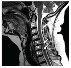
are studies that create better images of the body’s soft tissues. An MRI of the neck can show if your nerve compression is caused by damage to soft tissues—such as a bulging or herniated disk. It can also help your doctor determine whether there is any damage to your spinal cord or nerve roots.
This MRI image shows bulging disks pressing on the spinal cord (arrows).
TREATMENT OPTIONS
Nonsurgical Treatment
Initial treatment for cervical radiculopathy is nonsurgical. Nonsurgical treatment options include:
Soft cervical collar. This is a padded ring that wraps around the neck and is held in place with Velcro. Your doctor may advise you to wear a soft cervical collar to allow the muscles in your neck to rest and to limit neck motion. This can help decrease the pinching of the nerve roots that accompany movement of the neck. A soft collar should only be worn for a short period of time since long-term wear may decrease the strength of the muscles in your neck.
Physical therapy. Specific exercises can help relieve pain, strengthen neck muscles, and improve range of motion. In some cases, traction can be used to gently stretch the joints and muscles of the neck.
Medications. In some cases, medications can help improve your symptoms.
- Nonsteroidal anti-inflammatory drugs (NSAIDs). NSAIDs, including aspirin, ibuprofen, and naproxen, may provide relief if your pain is caused by nerve irritation or inflammation.
- Oral corticosteroids. A short course of oral corticosteroids may help relieve pain by reducing swelling and inflammation around the nerve.
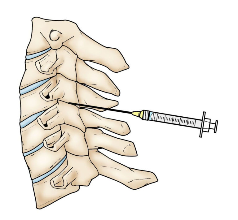
Facet joint injection in the cervical spine.
Steroid injection. In this procedure, steroids are injected near the affected nerve to reduce local inflammation. The injection may be placed between the laminae (epidural injection), in the foramen (selective nerve injection), or into the facet joint. Although steroid injections do not relieve the pressure on the nerve caused by a narrow foramen or by a bulging or herniated disk, they may lessen the swelling and relieve the pain long enough to allow the nerve to recover.
Narcotics. These medications are reserved for patients with severe pain that is not relieved by other options. Narcotics are usually prescribed for a limited time only.
Surgical Treatment
If after a period of time nonsurgical treatment does not relieve your symptoms, your doctor may recommend surgery. There are several surgical procedures to treat cervical radiculopathy. The procedure your doctor recommends will depend on many factors, including what symptoms you are experiencing and the location of the involved nerve root. Pre-Surgical EMG Testing is therefore critical to identify exactly what regions of the spine, canals and regional anatomy is affecting your nerve function.
GET RELIEF FROM PINCHED NERVES NOW
(312) 877-0035
Fast & Convenient
Expert EMG Neurological Diagnostic Tests - Same Day Testing Available! We ALSO Document work & Personal Injuries for legal cases as well. We have FOUR locations to serve you.